
FHO Surgery (Femoral Head Ostectomy)
Overview
What Is Femoral Head Ostectomy (FHO) Surgery In Dogs
FHO surgery is used when a dog’s hip joint has become a source of ongoing pain and is no longer working as a smooth, weight-bearing structure. Instead of trying to repair the joint, the procedure removes the part that is causing the problem.
In a healthy dog, the femoral head fits into the acetabulum to form a ball-and-socket joint. Cartilage covers both surfaces, and synovial fluid helps them glide smoothly so the hip can support weight and allow movement at the same time. When that system is damaged or unstable, the joint can no longer move cleanly, and each step can create friction, pressure, or bone-on-bone contact.
FHO removes the femoral head and neck so that painful contact is no longer possible. The joint itself is not rebuilt. Instead, the body adapts by using surrounding muscle and connective tissue to support movement in a different way. The goal is to reduce pain and allow the dog to use the limb more comfortably, even though the original joint structure is no longer there.
How The Hip Anatomy Changes In FHO Surgery
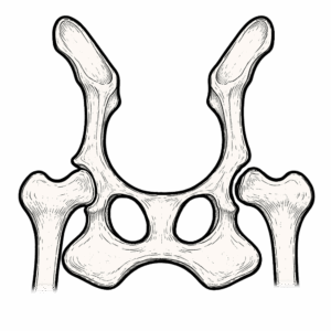
This chalkboard illustration diagram shows the key structural change of an FHO procedure. In the normal hip, the femoral head acts as the rounded ball of the joint and sits inside the acetabulum, the socket in the pelvis. After surgery, that ball is removed. The acetabulum remains, but it no longer receives a smooth femoral head, and the femur is no longer moving through a true ball-and-socket joint.

That change matters because the hip is no longer stabilized by normal joint architecture. The body heals the area with fibrous connective tissue, while the surrounding muscles and fascia take on a much greater role in holding the femur in a workable position and guiding movement. Fascia is the connective tissue network that surrounds and links muscles, helping them transfer force and work together. What develops is often called a pseudo-joint or false joint, but it is better understood as a functional adaptation than a replacement hip.
How The Hip Heals And Adapts After FHO Surgery
Immediately after surgery, the area contains post-operative inflammation, fluid, and disrupted tissue. As healing progresses, fibroblasts, cells that produce connective tissue, begin laying down collagen. Collagen is a structural protein that gives tissues strength. These fibers do not turn into cartilage and do not recreate the original joint surface. Instead, they form a dense fibrous interface between the upper femur and the pelvis.
At the same time, the muscles around the hip begin adapting to a different job. Before surgery, much of the hip’s stability came from the shape of the joint itself. After surgery, stability becomes much more dependent on active muscle support. This is why recovery is shaped not only by tissue healing, but by how the dog relearns movement while that tissue is organizing.
How Movement Works After FHO Surgery In Dogs
After FHO, the hip does not move like a normal joint, even in a strong recovery. In a healthy hip, the femoral head rotates within the socket in a relatively predictable pattern. After FHO, there is no true center of rotation. The femur is guided by soft tissue, especially the gluteal muscles, hamstrings, quadriceps, and the connective tissue that links them.
That changes how the limb bears weight, how the dog pushes off during walking, and how the nervous system senses the hip region. Early movement can look awkward even when the surgery itself was successful. The dog is not simply waiting for pain to fade. Muscles are relearning timing, connective tissue is organizing under load, and the nervous system is adjusting to a different mechanical pattern.
What Good Function Looks Like After FHO Surgery
A good result after FHO does not mean the hip has returned to normal. It means the dog can use the limb comfortably and reliably in daily life.
A dog with a strong functional recovery will usually:
- bear weight on the limb during walking
- rebuild muscle in the affected thigh
- move with a more even, repeatable gait
- rise, turn, and reposition with less hesitation
Some dogs still show subtle gait differences, especially at faster speeds or during harder activity. That alone does not mean the procedure failed. FHO is designed to improve comfort and usable function, not to recreate a normal hip joint.
Why Some Dogs Do Better With FHO Than Others
FHO can work very well, but outcomes are not identical across all dogs. Body size matters because a lighter dog places less force through the hip region with each step. Muscle condition matters because muscle becomes a major stabilizer after surgery. Age can matter because younger dogs often rebuild strength and adapt movement patterns more easily, while older dogs may take longer to regain confidence and coordination.
Activity level and willingness to use the limb also shape recovery. Dogs that begin engaging the leg and rebuilding strength often do better than dogs that remain highly protective of it. That is one reason active rehabilitation is so important to the final outcome.
When Veterinarians Recommend FHO Surgery
Veterinarians recommend FHO when the hip joint itself has become the source of ongoing pain and is no longer functioning in a way the body can use comfortably.
This most often develops over time. In hip dysplasia, the ball and socket do not fit tightly together. With each step, the femoral head shifts within the socket instead of staying centered. That repeated motion places uneven stress on cartilage and bone. The body responds with inflammation, tissue thickening, and structural changes that gradually make the joint stiffer and more painful to use.
Other conditions can reach a similar point through different mechanisms. A fracture of the femoral head or neck can disrupt the shape of the joint so it no longer moves smoothly. Chronic luxation prevents the femoral head from staying seated in the socket. In Legg-Calvé-Perthes disease, the bone of the femoral head weakens and begins to break down. In each case, the joint loses its ability to function as a stable, low-friction system.
At that stage, the question shifts from how to support the joint to whether the joint itself is still worth preserving.
In some dogs, earlier procedures can improve how the joint fits together, particularly when the problem is identified during growth. In others, total hip replacement may be considered to recreate the joint using implants and restore more typical mechanics.
FHO is chosen when those approaches are not the right fit for the situation. Instead of trying to preserve or replace the joint, it removes the part of the joint that is generating pain and allows the body to adapt around that change.
The timing of that decision can vary. Some veterinarians recommend surgery once it is clear the joint is driving pain and limiting function. Others may first use pain management, activity modification, and rehabilitation to see how much comfort can be restored without surgery.
The goal across approaches is the same. Reduce pain, and allow the dog to move in a way that is sustainable over time.
What Recovery Really Looks Like After FHO Surgery
Recovery after FHO is usually gradual. In the first several days, many dogs use the limb very little. Some toe-touch. Some hold the leg up. Some place it briefly and then pull away. That early stage often reflects post-operative soreness, caution, and the fact that the dog no longer has the same joint structure or movement pattern.
Then a more meaningful stage begins. The dog starts testing the limb. Weight-bearing appears in short bursts. Standing becomes more even. Toe contact becomes more regular. The leg is still weak, but the body is beginning to experiment with using it again.
As recovery progresses, the signs become more substantial:
- the dog uses the limb during slow walking more consistently
- the thigh begins to regain muscle
- push-off becomes more natural when rising or turning
- weight shifts become smoother and less guarded
When recovery stalls, the picture tends to look different. The limb stays thin, the dog continues shifting weight away from it, and the gait remains inconsistent. In many of these dogs, the problem is not that the incision did not heal. The problem is that the body settled into compensation instead of fully rebuilding limb use.
What The Immediate Post-Operative Period Looks Like After FHO Surgery
The first phase after FHO surgery is focused on healing, comfort, and early reintroduction of the limb. This period often feels uncertain because the dog is not yet moving in a way that looks improved.
After surgery, most dogs go home with a combination of pain management and activity restriction. Pain control is not just about comfort. It reduces protective muscle tension and makes it easier for the dog to begin using the limb as healing progresses.
Post-operative care commonly includes:
- anti-inflammatory medications to reduce tissue inflammation
- additional pain medications that act on the nervous system to reduce pain signaling
- activity restriction to prevent jumping, running, or sudden movements
- short, controlled leash walks for bathroom breaks and gentle movement
In the first several days, it is common for dogs to:
- hold the leg up or use it only briefly
- shift weight more heavily to the other limbs
- show lower energy or temporary changes in appetite
These responses reflect a combination of surgical soreness, protective behavior, and the fact that the hip no longer provides the same mechanical feedback it did before.
At the tissue level, healing is already underway. The surgical site contains inflammation, fluid, and early repair activity. The incision on the surface typically heals within 10 to 14 days, but deeper tissue remodeling continues well beyond that point.
As pain becomes better controlled and the dog begins to feel more stable, small changes start to appear. The dog may begin placing the toes on the ground while standing or using the leg briefly during slow walking. These early efforts are often inconsistent, but they mark an important shift. The limb is starting to be incorporated back into movement.
The goal during this phase is not normal movement. The goal is safe healing while keeping the limb involved. When a limb is used regularly, even in small amounts, it helps maintain muscle activity and gives the nervous system continued input from that side of the body. When use is very limited, muscle loss progresses and the dog begins relying more heavily on the other limbs, which can make later recovery more difficult.
This early phase sets the foundation for everything that follows. It is where healing begins, but also where the first movement patterns after surgery start to take shape.
What A Realistic FHO Recovery Timeline Looks Like
The first two weeks are mainly about incision healing, pain control, and preventing complete disuse. Dogs are usually not walking normally during this stage, but total inactivity is generally not the goal. Short, controlled movement helps keep the limb involved while healing begins.
From about two to six weeks, many dogs begin more deliberate use of the leg. Toe-touching may progress to partial weight-bearing during controlled walks. Day to day progress can look modest, but this period is important because movement habits start taking shape here.
From six to twelve weeks, muscle rebuilding and coordination become more visible. Dogs that are using the limb consistently often show a steadier gait, better push-off, and less guarded movement. Plateaus can happen here, especially once the early improvements level off and deeper strength-building is still underway.
Beyond three months, many dogs continue improving in endurance, confidence, and muscle mass. Some end up moving very well in everyday life, even if subtle differences remain during running, jumping, or high-demand activity. FHO recovery is best understood as a months-long adaptation process, not a short post-op event.
How Rehabilitation Shapes The Outcome After FHO Surgery
Rehabilitation matters because the tissues forming after surgery are shaped by use. Fibrous connective tissue develops between the femur and pelvis, muscles take on a larger stabilizing role, and the nervous system relearns how to load and control the limb. All of those processes respond to movement.
Controlled use of the leg gives the body useful information. It helps connective tissue align under stress, encourages muscle recruitment, and helps the brain and spinal cord keep incorporating the limb into normal movement. Without enough of that input, the area still heals, but it may heal into a less functional pattern.
Common rehabilitation strategies may include:
- controlled leash walking to build repeatable weight-bearing
- passive range-of-motion work to reduce stiffness and maintain mobility
- assisted standing or weight shifts to encourage early limb use
- hydrotherapy to support movement with less full body load
- progressive strengthening to rebuild muscle and coordination
- manual therapy to reduce guarding and improve tissue mobility
Different clinicians may emphasize different tools, but the goal is the same: help the dog build a stronger, more organized way of using the limb.
What Long-Term Success Looks Like After FHO Surgery
Long-term success after FHO means the dog is comfortable, uses the limb reliably, and can move through daily life without the hip remaining a major source of pain. The gait may not be perfectly symmetrical, and the limb may not function exactly like an untouched hip. That is not the standard FHO is trying to meet.
This procedure works best when expectations are realistic. It is not a joint restoration surgery. It is a functional salvage procedure that can produce very good quality of life when case selection is sound and recovery is supported well.
General Health Topics

Health Conditions

Follow the Research
| Title | Information |
|---|
Dig Deeper
| Title | URL | At a Glance |
|---|
Blog Articles
| Featured Image Link | Blog Title | Blog_URL_Link |
|---|