
Eyes & Vision
Overview
The Importance of Preventing Eye Issues
A dog’s eyes are remarkable organs of perception. They provide information about the environment, help coordinate movement, and play an essential role in how dogs connect with their world.
Healthy eyes allow a dog to navigate confidently, respond to subtle motion, and interpret visual cues that are vital for training and safety. When eyes become irritated, inflamed, or injured, a dog’s entire experience of the world changes. They may become hesitant to move, squint in discomfort, or withdraw from normal activities.
Eye problems are among the most common veterinary concerns, ranging from mild surface irritation to conditions that threaten vision. Because the eye’s structure is delicate and interconnected with multiple body systems, even small issues can quickly become serious if untreated. Understanding how the eye is built and how it functions helps pet parents recognize early warning signs and support lifelong ocular health.
Dog Eye Anatomy
The dog’s eye, or globe, is a precisely layered structure designed to capture and process light. It rests within a protective socket of bone and is cushioned by fat and connective tissue that allow smooth movement. While similar to the human eye in overall design, several features reflect adaptations to a dog’s unique visual needs.
The outermost layer includes the cornea, sclera, and conjunctiva. The cornea is the clear, curved surface that allows light to enter. It is extremely sensitive and must remain moist to function properly. The sclera, or white of the eye, provides structure and protection, while the conjunctiva lines the eyelids and front of the sclera, forming a barrier against debris and infection.
Beneath these layers lie the iris, pupil, and lens, which regulate and focus incoming light. The iris adjusts the size of the pupil depending on brightness, while the lens fine-tunes focus for near or distant vision. Behind the lens is the retina, a thin membrane containing specialized light-sensitive cells: rods for detecting motion and low light, and cones for detecting color and detail. The optic nerve transmits signals from the retina to the brain, where they are processed into recognizable images.
Dogs also possess a third eyelid, or nictitating membrane, located in the inner corner of the eye. This thin, semi-transparent structure sweeps across the eye to provide additional protection and lubrication. It is an important defense against debris and plays a role in tear production. The lacrimal glands, located above each eye, produce tears that keep the cornea hydrated and clean.
Together, these components allow the eye to maintain constant clarity, focus, and protection. Any disruption in this system—whether from dryness, inflammation, or injury—can impair vision or cause significant discomfort.
External Eye Anatomy Diagram: Front View
The front view of the dog’s eye highlights the external structures responsible for surface protection, tear production, and tear drainage. This perspective emphasizes features that are visible without magnification, such as the eyelids, third eyelid, sclera, and the structures of the lacrimal (tear) system. These components work together to maintain a healthy tear film, protect the eye from environmental debris, and regulate how tears flow across and away from the eye.

| Structure | Definition |
|---|---|
| Upper Eyelid | Protective upper skin fold that distributes tears and shields the eye. |
| Lacrimal Caruncle | Small pink mound at the inner corner of the eye containing glands that help produce components of the tear film. |
| Lacrimal Canaliculi | Small drainage channels that collect tears from the eye’s surface and carry them toward the lacrimal sac. |
| Nasolacrimal Duct | Passage that drains tears from the lacrimal sac into the nasal cavity. |
| Third Eyelid (Nictitating Membrane) | Inner protective eyelid that sweeps across the eye to clear debris and support tear production. |
| Iris | Colored portion of the eye that adjusts the size of the pupil to regulate light entry. |
| Pupil | Central opening in the iris that allows light to enter the eye. |
| Sclera | White, fibrous outer layer that maintains eye shape and provides protection. |
| Tarsal Gland Openings (Lower Tear Ducts / Meibomian Glands) | Openings of oil-producing glands along the eyelid margins that stabilize the tear film and reduce evaporation. |
Internal Eye Anatomy Diagram: Side Cross-Section View
This side view, or cross-section, reveals the internal anatomy that enables vision, including the cornea, lens, retina, and supporting tissues that guide and process light. This deeper perspective shows how the outer protective structures connect to the eye’s internal chambers and how light passes through transparent tissues before being converted into neural signals.

| Structure | Definition |
|---|---|
| Upper Eyelid | Upper skin fold that protects the eye and spreads tears evenly over the cornea. |
| Lower Eyelid | Lower skin fold that supports protection and tear film distribution. |
| Third Eyelid | Inner eyelid that moves across the eye for protection and lubrication. |
| Cornea | Clear, curved front surface of the eye providing most of the eye’s focusing power. |
| Iris | Pigmented muscular structure that controls the size of the pupil. |
| Pupil | Opening in the center of the iris that determines how much light enters the eye. |
| Lens | Transparent, flexible structure that changes shape to focus light onto the retina. |
| Sclera | Tough outer coat of the eyeball, maintaining structure and protecting internal components. |
| Retina | Light-sensitive inner layer that converts light into neural signals sent to the brain. |
| Blood Vessels of the Retina | Network of vessels supplying oxygen and nutrients to the retina. |
How Vision Works for Dogs
Vision begins when light enters the cornea and passes through the pupil to reach the lens. The lens bends, or refracts, the light to focus it onto the retina. The retina contains millions of photoreceptor cells that convert light into electrical signals. These signals travel along the optic nerve to the brain’s visual cortex, where they are interpreted as shapes, colors, and motion.
Dogs rely heavily on motion detection rather than fine detail. Their eyes are adapted for low-light vision, thanks to a high concentration of rod cells and a reflective layer behind the retina called the tapetum lucidum. This layer acts like a mirror, amplifying available light and giving dogs superior night vision. It is also responsible for the familiar glow of a dog’s eyes when illuminated in the dark.
Depth perception and field of view are influenced by the placement of the eyes on the head. Most dogs have a wide field of vision, typically around 240 degrees, compared to about 180 degrees in humans. However, their binocular overlap—the area where both eyes’ visual fields intersect—is smaller, meaning they perceive less fine detail.
Although dogs do not see the full color spectrum humans do, they are not colorblind. They see primarily in shades of blue and yellow, with limited perception of red and green. Their visual system prioritizes motion and contrast, which is ideal for detecting prey or subtle body movements.
The eye’s intricate coordination with the brain and surrounding muscles ensures constant adjustment to light, focus, and movement. This system allows dogs to interpret their surroundings efficiently and stay oriented in changing conditions.
Why Dogs Are Vulnerable to Eye Issues
Dogs’ eyes evolved to support survival behaviors such as tracking movement, navigating in low light, and surveying wide areas. These adaptations give dogs meaningful advantages, but they also create structural vulnerabilities.
A dog’s eye shape plays a major role in what kinds of issues they are likely to develop. Breeds with loose or heavy eyelids, including cocker spaniels and many bulldog types, can put extra strain on the tissues that hold the third-eyelid gland in place. When this gland slips forward and becomes visible as a pink mass in the inner corner of the eye, it creates a condition known as cherry eye. Loose eyelids can also affect how tears spread across the eye, contributing to dryness or irritation.
Breeds with prominent or protruding eyes, such as pugs and Shih Tzus, face different risks. Their eyes sit farther forward and have less natural protection from the eyelids and surrounding bone. This makes them more prone to dryness, scratches, accidental trauma, and surface infections.
Dogs with deep-set or tighter eyelids may experience inward- or outward-rolling lids that cause chronic tearing, redness, or irritation. Across all breeds, the natural shape of the eye—whether protruding, loose-lidded, or recessed—strongly influences how easily problems can develop.
Dogs also rely on a third eyelid for protection and tear production. While this structure is helpful, it can become irritated or swollen like any other tissue. The tear film that coats the cornea is another key defense; when tear production decreases—as in dry eye (keratoconjunctivitis sicca)—the cornea becomes more vulnerable to injury and infection.
Because the eye’s tissues are delicate and react quickly to irritation, problems can worsen rapidly. Allergies, infections, or systemic diseases often show up first as redness, squinting, cloudiness, or discharge. Noticing these signs early—and addressing them promptly—is important for protecting a dog’s long-term comfort and vision.
Common Dog Eye Problems
Dog eye problems are among the most common reasons pet parents search for help, especially when a dog’s eyes suddenly look “weird,” cloudy, red, watery, swollen, or irritated. Many of these conditions start mild but can progress quickly because the eye is such a delicate, specialized organ.
Conjunctivitis (Pink Eye in Dogs)
Dogs can develop conjunctivitis, commonly called pink eye. Conjunctivitis occurs when the conjunctiva—the thin, clear tissue lining the eyelids and covering part of the eye—becomes inflamed. This inflammation can be caused by allergies, bacterial or viral infections, irritants like dust or smoke, or problems with tear drainage, such as a blocked tear duct.
Common signs include redness of the eye, frequent blinking or squinting, swelling of the eyelids, and discharge. The discharge may be watery or thick and mucus-like, often forming crusts around the eyes, especially after sleep. Symptoms can affect one or both eyes and may range from mild irritation to more noticeable discomfort.
Eye Infections
Eye infections can affect the surface of the eye or the tissues around it. They may develop from bacteria, viruses, fungi, or foreign debris. Symptoms often include redness, thick yellow or green discharge, swelling, and sensitivity to light. Infections can occur on their own or as a complication of pink eye, dry eye, or an injury.
Dry Eye (Keratoconjunctivitis Sicca)
Dry eye occurs when tear production drops and the surface of the eye no longer stays properly coated. Without enough tears, the cornea becomes irritated, sticky, or cloudy. Dogs may develop thick discharge, eye infections, or painful ulcers.
Cherry Eye
Cherry eye is the visible bulging of the third-eyelid gland in the inner corner of the eye. It appears as a red or pink mass. While not usually an emergency, the exposed gland can become inflamed or infected and often needs surgical correction to restore normal tear production.
Corneal Ulcers and Scratches
The cornea is extremely sensitive. A scratch from play, debris, or rubbing the face can quickly turn into a corneal ulcer. Signs include squinting, pawing at the face, excessive tearing, and sensitivity to light.
Eyelid Problems (Entropion, Ectropion)
Some dogs are born with eyelids that roll inward (entropion) or outward (ectropion), and certain breeds may even develop both conditions in the same dog, though they usually affect different eyelids.
- Entropion causes the eyelid margin and eyelashes to turn inward, rubbing against the cornea and causing pain, tearing, and irritation.
- Ectropion creates a loose, droopy eyelid that leaves the eye more exposed and prone to dryness, debris, and infection.
Both conditions can lead to redness, chronic tearing, irritation, and recurrent eye problems if not treated.
Cataracts
Cataracts create a white, opaque area in the lens that blocks light and can cause partial or full blindness. They develop from genetics, diabetes, aging, or injury. Cataracts are often confused with another age-related change—nuclear sclerosis.
Nuclear Sclerosis (Not Cataracts)
Nuclear sclerosis is a normal aging change where the lens becomes bluish or cloudy but stays transparent. Dogs with nuclear sclerosis can still see well. In contrast, cataracts block light and impair vision. This distinction matters because many pet parents worry their “old dog eyes” mean disease when the change may be harmless.
Glaucoma (High Eye Pressure)
Glaucoma occurs when pressure builds inside the eye and damages the optic nerve. It is extremely painful and can cause rapid, irreversible blindness. Symptoms include a swollen or enlarged eye, cloudy cornea, redness, and sudden behavioral changes. High eye pressure is a true emergency.
Progressive Retinal Atrophy (PRA)
PRA is a genetic disease that slowly damages the retina. Dogs often lose night vision first, then peripheral vision, and eventually become fully blind. There is no cure, but early diagnosis helps owners adapt the home to keep blind dogs safe and confident.
Tear Stains
Tear stains happen when tears overflow onto the fur and oxidize, leaving reddish-brown streaks. They can be caused by anatomy (shallow eye sockets), blocked tear ducts, allergies, or tear overproduction. Breeds like poodles and chihuahuas are commonly affected.
When to See a Veterinarian for Eye Issues
Because dog eye conditions can worsen quickly, it’s important to know when a change in your dog’s eyes is a minor irritation and when it signals a more serious problem. A veterinarian can perform a focused eye exam that evaluates the surface of the eye, tear production, internal pressure, and the deeper structures responsible for vision.
Seek veterinary care promptly if your dog has:
- Redness, swelling, or squinting
- Sudden tearing, discharge, or excessive “eye boogers”
- A cloudy, bluish, or milky-looking eye
- A visible red or pink lump in the inner corner (possible cherry eye)
- Eyelids that roll inward or outward
- One eye that suddenly looks “weird” or different from the other
- Trouble seeing in dim light or signs of partial blindness
- Rubbing or pawing at the face
- A bad odor coming from the eye
- A change in pupil size or eye shape
These signs suggest the eye needs attention before damage worsens.
Eye Problems That May Require an Emergency Visit
Some problems are urgent and should be seen the same day:
- Sudden blindness or disorientation
- A bulging eye (can signal glaucoma or trauma)
- Severe squinting or obvious pain
- Blood inside or around the eye
- Rapid clouding of the cornea
- A deep scratch or ulcer visible on the eye surface
Eye emergencies can progress rapidly, and early care can save vision.
When to See a Canine Ophthalmologist (Eye Specialist)
Most eye problems can be handled by your regular veterinarian, but some conditions need the training and equipment of a board-certified veterinary ophthalmologist. These specialists diagnose and treat complex eye diseases and perform delicate procedures that help protect or restore vision.
Your dog may need an ophthalmologist if they have:
- Glaucoma or dangerously high eye pressure
- Cataracts or possible cataract surgery
- Non-healing corneal ulcers
- Cherry eye that requires repair
- Eyelid problems (entropion, ectropion, or eyelid tumors)
- Suspected retinal disease or unexplained vision loss
- Severe or complicated trauma to the eye
These conditions involve deeper structures of the eye or require precise treatment.
A canine ophthalmologist can:
- Use advanced imaging to examine the retina, optic nerve, and lens
- Measure eye pressure with high-accuracy tools for glaucoma diagnosis
- Perform microsurgery, including cataract removal and corneal repairs
- Create specialized treatment plans for long-term conditions like autoimmune dry eye or chronic inflammation
What Happens During a Dog Eye Exam
A complete veterinary eye exam looks at the surface, structures, and function of the eye. Several simple, non-painful tests are commonly used:
| Test / Procedure | What It Is | What It Detects | Why It Matters |
|---|---|---|---|
| Fluorescein Stain Test | A drop of fluorescent dye is placed on the eye and rinsed; damaged areas glow under blue light. | Corneal scratches, ulcers, worn areas on the surface. | Ulcers can worsen quickly and may require immediate treatment to prevent infection or scarring. |
| Schirmer Tear Test | A small paper strip is placed under the lower eyelid for one minute to measure tear production. | Dry eye (keratoconjunctivitis sicca, KCS). | Low tear production leads to irritation, infection, and corneal damage if untreated. |
| Tonometry | A handheld tool briefly touches the cornea to measure intraocular pressure (IOP); quick and painless. | Glaucoma (high pressure) or uveitis (low pressure). | High eye pressure is an emergency that can cause rapid, permanent blindness. |
| Slit-Lamp Biomicroscopy | A bright, focused light and magnifier allow close examination of eye structures. | Cataracts, inflammation, eyelid issues, corneal defects, tumors. | Helps evaluate deeper structures with precision, often before surface signs appear. |
| Pupil Dilation & Retinal Exam | Eye drops dilate the pupil so the inner eye can be viewed with a special light. | Cataracts, retinal disease, optic nerve issues, PRA signs. | Critical for diagnosing internal problems that affect vision long-term. |
| Ocular Ultrasound / Imaging | Sound waves or imaging tools visualize structures when the inside of the eye is cloudy. | Retinal detachment, bleeding, masses, lens dislocation. | Allows diagnosis even when the view into the eye is blocked by cataracts or swelling. |
Eye Care Tips for Dogs
Routine observation and gentle care are the foundations of eye health. Pet parents should check their dog’s eyes regularly for redness, discharge, cloudiness, tearing, or changes in pupil size. Healthy eyes are clear, moist, and free of crust. The area around the eyes should stay clean, especially in dogs with wrinkles or long facial hair, where moisture can collect.
Clean the outer corners of the eyes with a soft, damp cloth or a veterinary-approved wipe. Avoid touching the cornea itself, as it is extremely sensitive. Dogs with light coats may develop tear staining; regular grooming, good tear drainage, and keeping the area dry can reduce discoloration and skin irritation.
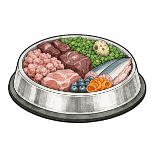
A balanced diet supports eye health from the inside out. Nutrients such as omega-3 fatty acids, vitamin A, lutein, and antioxidants help protect the retina and keep the tear film stable. Proper hydration and overall immune balance also contribute to healthy eyes and clearer vision.
Age-Related Eye Changes
As dogs grow older, their eyes naturally change. A common example is nuclear sclerosis, a bluish haze in the lens that does not block vision. Senior dogs may also develop reduced night vision, slower pupil responses, and a higher risk of cataracts or retinal disease. Regular veterinary exams become increasingly important with age, since early detection helps distinguish normal aging from true disease.
Environmental and Lifestyle Care
Environmental management can prevent irritation. Limiting exposure to dust, smoke, wind, or harsh cleaning chemicals reduces strain on the surface of the eye. When walking or riding in open vehicles, keeping your dog’s head inside and protected helps prevent scratches and debris injuries.
Regular veterinary visits allow for early identification of conditions like glaucoma, dry eye, or cataracts—diseases that may progress quietly before obvious symptoms appear.
Simple Home Adaptations for Vision Changes
Dogs with reduced or fading vision can still live safe, confident, and comfortable lives with small adjustments at home:
- Keep furniture and walkways consistent to help with navigation.
- Use textured rugs to create “pathways” between key areas like food, water, and sleeping spots.
- Block off stairs or high ledges for dogs with depth-perception issues.
- Add night-lights in hallways to support senior dogs with declining low-light vision.
- Use voice cues or gentle sounds to help guide a dog who can no longer rely on visual cues.
- Avoid rearranging furniture if your dog is adapting to blindness or PRA.
Small changes make a big difference. With routine care, good nutrition, and attentive observation, pet parents can preserve both comfort and vision throughout a dog’s life.
The Eye in the Bigger Picture of Health
The eye is closely tied to the broader systems of the body. It is lined with mucous membranes, supplied with fine blood vessels, and influenced by immune and neurological activity. Many systemic conditions—such as diabetes, hypertension, autoimmune disease, or hormonal imbalance—can first show symptoms in the eyes.
Because the cornea, lens, and retina are sensitive to inflammation, recurring eye issues may signal deeper imbalances within the body. The immune system, in particular, plays a role in regulating inflammation and healing within the eye. Allergies and nutritional deficiencies can also disrupt normal function.
Maintaining ocular health, therefore, extends beyond the surface of the eye. It reflects the dog’s internal health, diet, hydration, and overall metabolic stability. Protecting vision means supporting every system that keeps the body in equilibrium.
Health Conditions
| Image & Title | At a Glance |
|---|---|

|
Entropion is a condition where a dog's eyelid rolls inward, causing the eyelashes and skin to rub against the eye, leading to irritation, pain, and potential corneal damage. It is most commonly seen in certain breeds with loose facial skin and is typically corrected with surgery. Supportive care, such as lubricating eye drops and pain management, may help relieve discomfort before and after surgical treatment. In a healthy eye, the eyelids sit smoothly against the globe and protect it; with entropion, that protective structure becomes a source of ongoing mechanical injury. Early recognition and correction are important to preserve comfort and long-term eye health. |

|
Cherry eye in dogs, medically called prolapsed gland of the third eyelid or nictitating membrane gland prolapse, occurs when a tear-producing gland in the inner corner of the eye slips out of its normal position. It appears as a pink or red bulge and is most common in young dogs. Because this gland produces a significant portion of the eye’s watery tears, displacement can affect long-term tear balance and ocular surface health. Left unaddressed, chronic exposure and inflammation can increase the risk of dry eye and ongoing irritation. |

|
Ectropion in dogs is a structural eyelid condition in which the lower eyelid rolls outward, exposing the inner lining of the eye. This outward turning disrupts normal tear distribution and leaves delicate tissues vulnerable to dryness, debris, and chronic irritation. While some breeds are predisposed due to facial conformation, significant ectropion can lead to recurrent conjunctivitis and long-term surface inflammation. Early evaluation helps determine whether supportive care is sufficient or surgical correction is needed. |
Therapeutic Interventions

Lifestyle Strategies
Foods
Food Components
Food Component Groups
Nutrients


Nutrient Types
Nutrient Sub-Types
Follow the Research
| Title | Information |
|---|
Dig Deeper
Blog Articles
| Featured Image Link | Blog Title | Blog_URL_Link |
|---|---|---|
 |
Brown Eyes In Dogs--Why Are They Dominant? | https://www.bernies.com/blogs/bernies-blog/brown-eyes-in-dogs-why-are-they-dominant/ |

|
The Eyes Have It | https://www.bernies.com/the-eyes-have-it/ |

|
The Connection Between Omega-3 and Your Dog’s Eye Health | https://www.bernies.com/blogs/bernies-blog/the-connection-between-omega-3-and-your-dogs-eye-health/ |